COLLABORATIVE ADVANCED RESEARCH IMAGING (CARI)
Collaborative Advanced Research Imaging (CARI)
For general inquiries about Collaborative Advanced Research Imaging (CARI) resources, please contact:
- Robert Cadrain
- 1 (804) 828-3639
- cctr-cari@vcu.edu
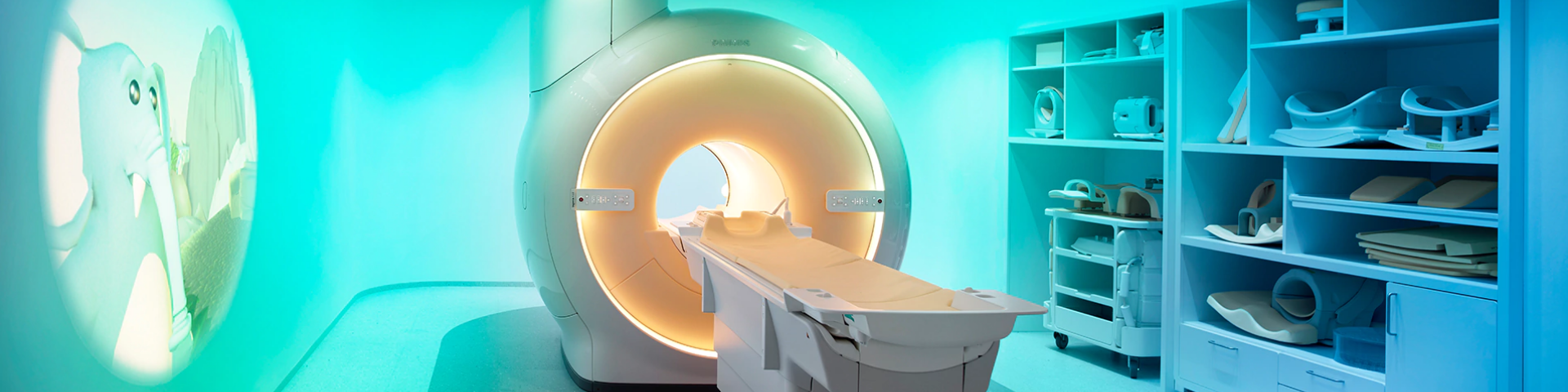
Our Collaborative Advanced Research Imaging (CARI) Center houses 6,000 square feet of research space including our research-dedicated Philips Ingenia 3.0 Tesla MRI Scanner, a mock scanner, interview rooms, drug screening rooms, a dispensary and more.
CARI MRI
- Uses Philips' Direct Digital processing, which substantially improves the signal-to-noise ratio
- Includes both front and rear-facing mirrors for visual stimuli and movie projection
- The radiofrequency signal is totally digital at the source in order to reduce noise
- Patient-specific B0 and B1 shimming
- Large bore to help participants feel less confined and less claustrophobic
In addition to the research-dedicated scanner, the CARI facility has more than 6,000 square feet of research-dedicated space, which houses the following:
- Mock scanner
- Includes both front and rear-facing mirrors for visual stimuli and movie projection
- Urine drug screen collection toilet
- Multiple interview rooms
- Sound attenuated testing chambers for human behavioral laboratory testing
- Physical exam room with electrocardiogram
- Dispensary for medication storage and dispensing
Upgrades to the CARI research dedicated MRI Scanner include MR shear Elastography (MRE) to measure liver fibrosis by automatically generating quantitative images (elastograms) of the entire liver that depict shear stiffness in units of kilopascals (kPa).
We also have Cardiac Expert Specialist for acquisition of multi-slice, dynamic tissue studies with saturation prepulse (for T1 weighting), WET saturation pulses (B1 insensitive) for uniform tissue suppression on 3.0T, Look Locker methods for determination of optimal inversion delay time, myocardial tagging with REST grids for regional wall motion studies, and real-time interactive imaging. Another recent cardiac MRI upgrade is StarQuant which provides single breathhold, multi-echo, ECG-triggered acquisitions to provide T2*, R2*, T2 and R2 maps for assessment of myocardial tissue characteristics.
Most recent upgrades to the research-dedicated MRI Scanner include the T2-Dixon pulse sequence, which acquires 4 image types in one single TSE scan (water, in-phase, out-of-phase, and fat) and provides uniform and consistent fat-free imaging, over large field of views and in challenging anatomies such as head, neck, spine, and muscular-skeletal. Another upgrade is the Philips MR400 Expression Patient Monitor, which monitors Blood Pressure, ECG, Pulse-ox SpO2, End-tidal CO2, and Body Temperature, while patients are being scanned in real-time in the Philips MRI scanner, and the physiological signals are synchronized with the scans and are recorded electronically by the scanner.
Please note that CARI is a research dedicated MRI facility. Results may not be used for clinical or diagnostic purposes. As such, all costs incurred must be study billed and are not eligible for third-party payments/billing/reimbursement. Please note that CARI is not affiliated with VCUHS or the VCU Department of Radiology.
To request CARI services, please click the link below. Thank you for your interest in CARI (Collaborative Advanced Research Imaging), located at 203 E Cary Street.
Request CARI services here
Instructions on how to submit for a quote may be found in our SOPs
Note to External Clients (external to VCU)– please select “CARI Services Only” when completing the in-take form. You will not be required to complete additional sections for required for VCU based studies.
CARI is distinguished as the only research-dedicated large bore MRI at VCU, providing unparalleled imaging capabilities specifically for research purposes. This exclusive focus on research allows us to tailor our services to meet the complex needs of investigators, ensuring that all imaging is performed in compliance with rigorous study protocols and federal regulations.
Results from CARI MRI may not be used for clinical or diagnostic purposes, and all costs incurred must be study billed, without eligibility for third-party payments or billing, including Standard of Care or Routine Care charges.